QUESTION IMAGE
Question
activity 17 : quiz
module : nervous
topic : structures / type : combination
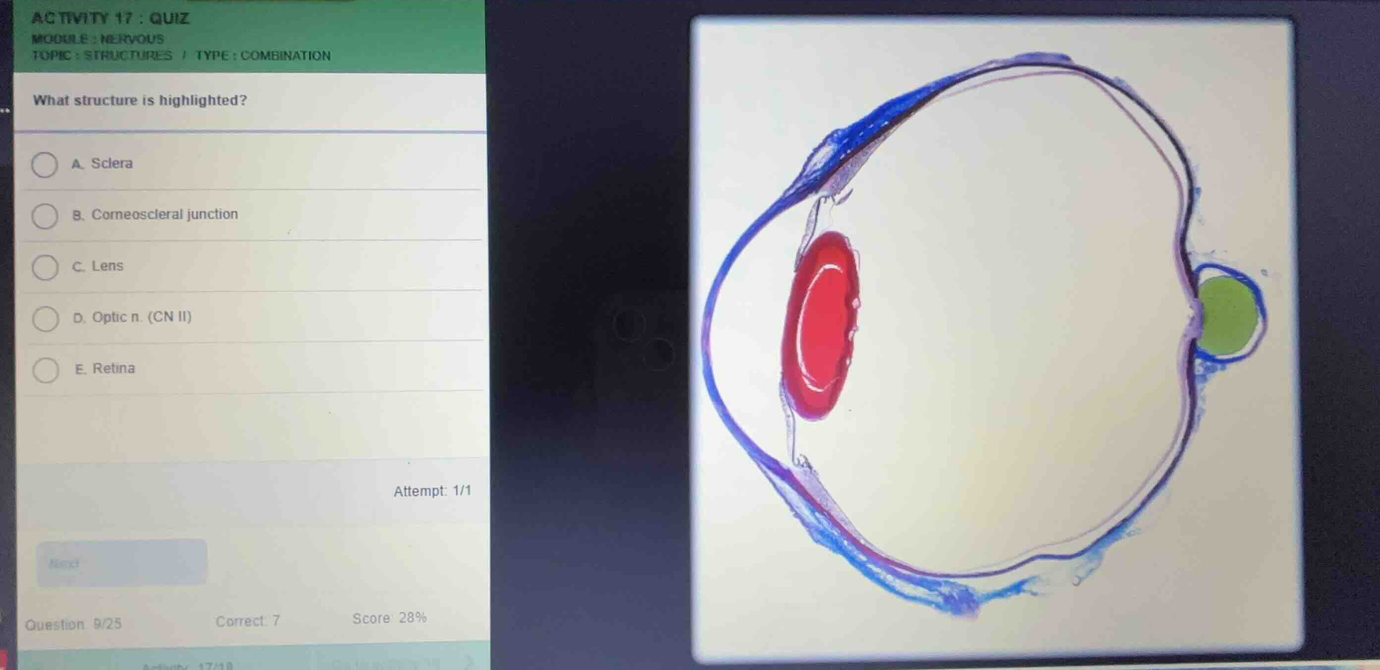
what structure is highlighted?
a. sclera
b. corneoscleral junction
c. lens
d. optic n. (cn ii)
e. retina
Identify the highlighted structure
Using the Eye Anatomy knowledge point
The histological section of the eye shows several color-coded regions:
- The lens is highlighted in red.
- The optic nerve (CN II) exiting the posterior aspect of the eye is highlighted in green.
- A thin inner layer along the posterior cavity is highlighted in pink/purple.
The question asks to identify the highlighted structure. Looking closely at the green highlighted region at the back of the eye, it corresponds to the exiting cranial nerve.
Correlate with anatomical structures
Using the Retina Anatomy knowledge point
- Sclera: The outer fibrous protective layer of the eye (shown in blue on the periphery).
- Corneoscleral junction: The transition zone between the cornea and sclera at the anterior eye.
- Lens: The biconvex structure suspended behind the iris (highlighted in red).
- Optic n. (CN II): The nerve bundle exiting the posterior pole of the eyeball (highlighted in green).
- Retina: The innermost nervous tissue layer lining the posterior eye (highlighted with a thin pink line).
Since the green highlighted structure is the nerve bundle exiting the posterior eye, it is the optic nerve.
Select the correct option
The green highlighted structure matches option D, Optic n. (CN II).

Snap & solve any problem in the app
Get step-by-step solutions on Sovi AI
Photo-based solutions with guided steps
Explore more problems and detailed explanations
- (A) Sclera
- (B) Corneoscleral junction
- (C) Lens
- (D) Optic n. (CN II) (Correct answer)
- (E) Retina