QUESTION IMAGE
Question
activity 17 : quiz
module : nervous
topic : structures / type : combination
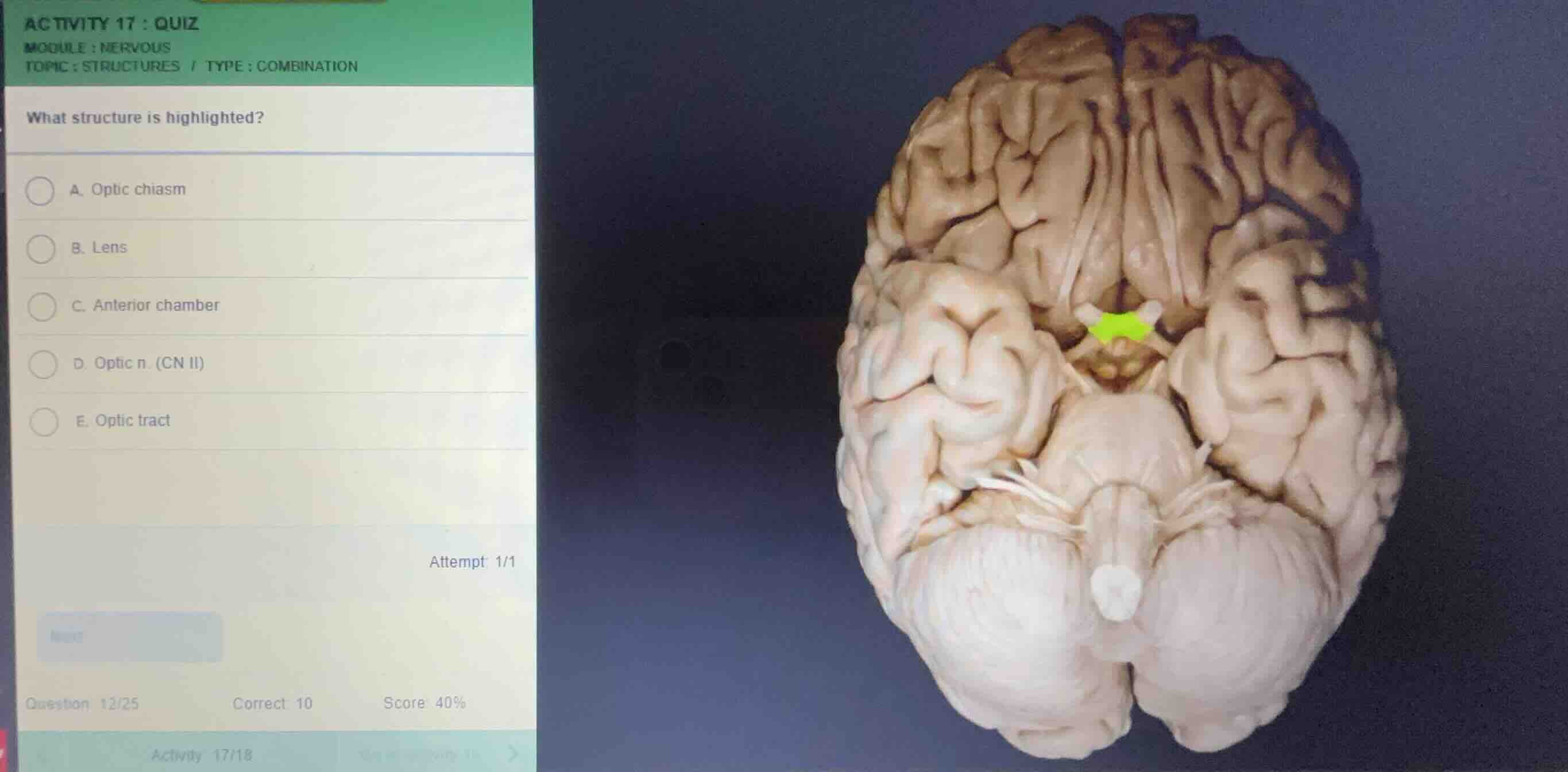
what structure is highlighted?
a. optic chiasm
b. lens
c. anterior chamber
d. optic n. (cn ii)
e. optic tract
attempt 1/1
question 12/25
correct 10
score 40%
Identify the view of the brain specimen
The image shows an inferior (ventral) view of a human brain specimen, with the anterior portion (frontal lobes) at the top and the posterior portion (cerebellum and brainstem) at the bottom.
Analyze the highlighted structure
Using the Brain Anatomy knowledge point, we locate the highlighted green region situated at the midline, just anterior to the pituitary stalk (infundibulum) and hypothalamus. This X-shaped structure is where the two optic nerves meet and cross.
Differentiate between the anatomical options
- The optic nerves (CN II) are the anterior bundles leading into this junction.
- The optic tracts are the posterior bundles continuing from this junction toward the brain.
- The central X-shaped intersection itself is the optic chiasm.

Snap & solve any problem in the app
Get step-by-step solutions on Sovi AI
Photo-based solutions with guided steps
Explore more problems and detailed explanations
- A. Optic chiasm (Correct answer)
- B. Lens
- C. Anterior chamber
- D. Optic n. (CN II)
- E. Optic tract