QUESTION IMAGE
Question
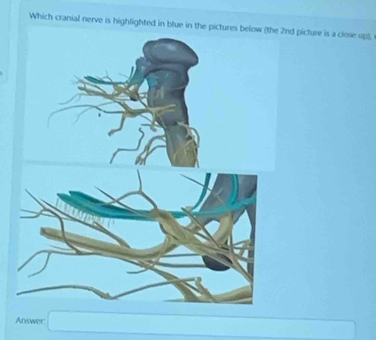
which cranial nerve is highlighted in blue in the pictures below (the 2nd picture is a close up),
Identify anatomical landmarks
The image displays the brainstem (pons and medulla) along with emerging cranial nerves. The highlighted structure in blue is a very thick nerve emerging from the lateral aspect of the pons.
Analyze nerve branches
The close-up image shows this thick nerve trunk splitting into three major branches: the ophthalmic (\(\text{CN V}_1\)), maxillary (\(\text{CN V}_2\)), and mandibular (\(\text{CN V}_3\)) divisions. This trifurcation is the classic signature of the trigeminal nerve.
Correlate with cranial nerve numbering
The trigeminal nerve is the fifth cranial nerve, designated as CN V. It is the largest of the cranial nerves and is responsible for sensation in the face and motor functions such as biting and chewing.

Snap & solve any problem in the app
Get step-by-step solutions on Sovi AI
Photo-based solutions with guided steps
Explore more problems and detailed explanations
Which cranial nerve is highlighted in blue in the pictures below (the 2nd picture is a close up) <blank>Trigeminal nerve</blank>