QUESTION IMAGE
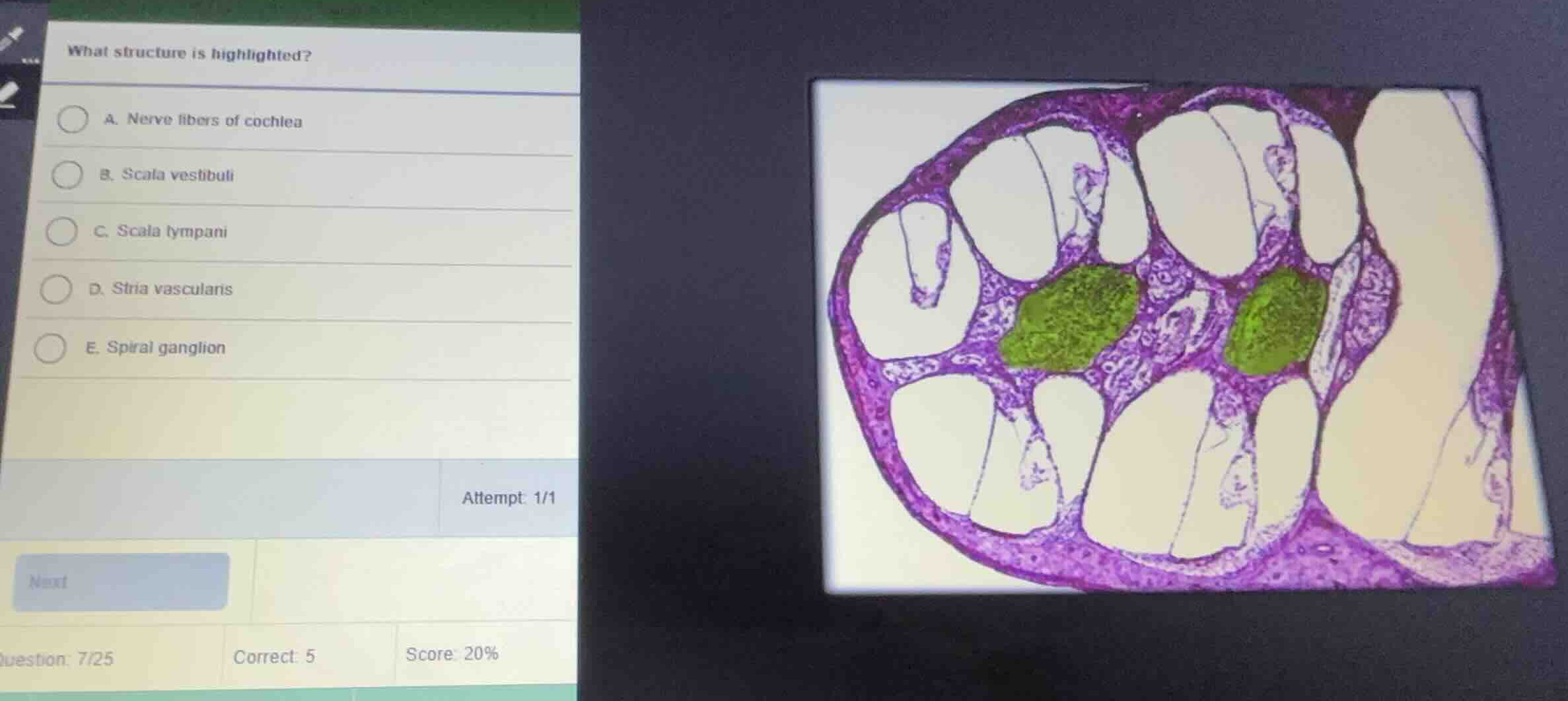
Question
what structure is highlighted?
a. nerve fibers of cochlea
b. scala vestibuli
c. scala tympani
d. stria vascularis
e. spiral ganglion
Identify the histological section
Using the Cochlea Histology knowledge point
The image shows a cross-section of the bony cochlea of the inner ear, which spirals around a central bony core known as the modiolus.
Analyze the highlighted structure
The green highlighted regions are located within the modiolus (the central axis of the cochlea). These clusters contain the cell bodies of the bipolar sensory neurons that innervate the hair cells of the organ of Corti. These cell bodies form the spiral ganglion.
Differentiate from other options
- Nerve fibers of cochlea (Option A) are the axonal projections traveling centrally from the ganglion through the modiolus.
- Scala vestibuli (Option B) and Scala tympani (Option C) are fluid-filled chambers within the cochlear turns.
- Stria vascularis (Option D) is located on the outer wall of the cochlear duct.
- Therefore, the highlighted clusters of neuronal cell bodies represent the Spiral ganglion (Option E).

Snap & solve any problem in the app
Get step-by-step solutions on Sovi AI
Photo-based solutions with guided steps
Explore more problems and detailed explanations
- A. Nerve fibers of cochlea
- B. Scala vestibuli
- C. Scala tympani
- D. Stria vascularis
- E. Spiral ganglion (Correct answer)