QUESTION IMAGE
Question
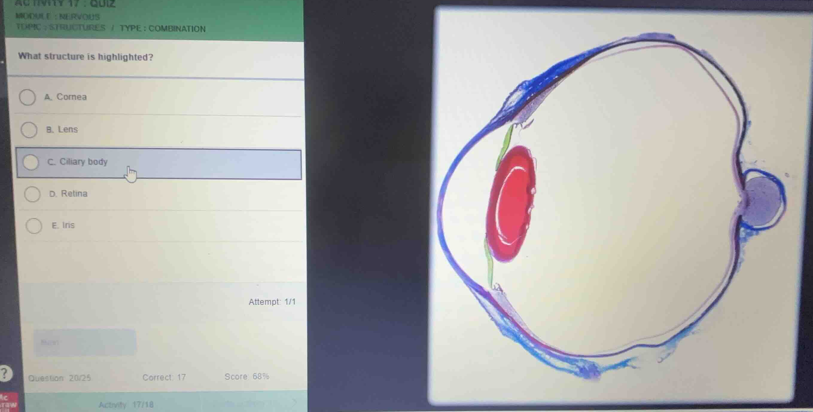
what structure is highlighted?
a. cornea
b. lens
c. ciliary body
d. retina
e. iris
Identify the highlighted structure in the histological section
Using the Eye Anatomy and Ocular Histology knowledge points, we analyze the sagittal cross-section of the eye. The large, oval, biconvex structure located in the anterior segment of the eye, colored bright red in this stained histological diagram, is the lens.
Analyze the anatomical position and features of the lens
Using the Lens Structure knowledge point, we confirm that the highlighted red structure is positioned directly behind the iris (colored green) and pupil, suspended by zonule fibers. This biconvex structure is responsible for focusing light onto the retina at the back of the eye.
Evaluate the multiple-choice options
- A. Cornea: The clear, outermost anterior layer (colored blue on the far left).
- B. Lens: The highlighted bright red biconvex structure.
- C. Ciliary body: The thickened portion of the vascular tunic connecting the choroid with the iris (near the base of the green iris).
- D. Retina: The inner neural layer lining the back of the eye (colored pink/purple on the inner posterior wall).
- E. Iris: The colored contractile diaphragm (colored green, anterior to the lens).

Snap & solve any problem in the app
Get step-by-step solutions on Sovi AI
Photo-based solutions with guided steps
Explore more problems and detailed explanations
- (A) Cornea
- (B) Lens (Correct answer)
- (C) Ciliary body
- (D) Retina
- (E) Iris