QUESTION IMAGE
Question
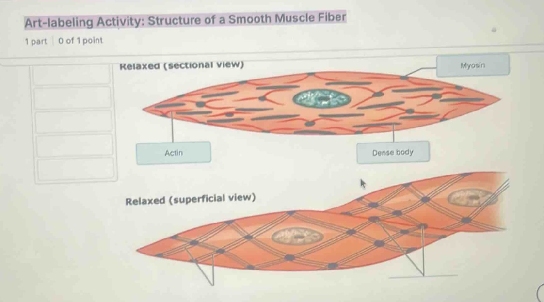
art-labeling activity: structure of a smooth muscle fiber
relaxed (sectional view)
myosin
actin
dense body
relaxed (superficial view)
🆕 New Concept Discovered: Smooth Muscle Microanatomy
How internal contractile proteins organize without sarcomeres.
Step 1: Identify the structure of smooth muscle fibers
Unlike skeletal muscle, smooth muscle does not contain highly organized sarcomeres (which give skeletal muscle its striped or striated appearance). Instead, contractile proteins are arranged in a loose, lattice-like network throughout the sarcoplasm:
- Actin (thin filaments): Attached to anchoring structures.
- Myosin (thick filaments): Interspersed between the actin filaments to allow sliding and contraction.
- Dense bodies: Functionally analogous to the Z-discs of skeletal muscle. They anchor the thin (actin) filaments and intermediate filaments both within the sarcoplasm and to the sarcolemma (cell membrane).
Step 2: Analyze the superficial view labels
In the bottom diagram labeled Relaxed (superficial view), we see the external lattice network wrapping around the spindle-shaped smooth muscle cell:
- Bottom-left pointer: Points to the network of thin lines running diagonally across the surface of the cell. These represent the Intermediate filaments (or non-contractile cytoskeletal elements/actin filament bundles associated with the membrane).
- Bottom-right pointer: Points directly to the dark, circular nodes where these filaments intersect on the cell membrane. These surface anchoring points are Dense bodies (specifically, membrane-associated dense plaques/bodies).

Snap & solve any problem in the app
Get step-by-step solutions on Sovi AI
Photo-based solutions with guided steps
Explore more problems and detailed explanations
Based on standard anatomical labeling for the Relaxed (superficial view) of a smooth muscle fiber:
- Bottom-left label: Intermediate filaments (or Actin/myofibril bundles)
- Bottom-right label: Dense bodies (or Dense plaques)