QUESTION IMAGE
Question
activity 17: quiz
module: nervous
topic: structures / type: combination
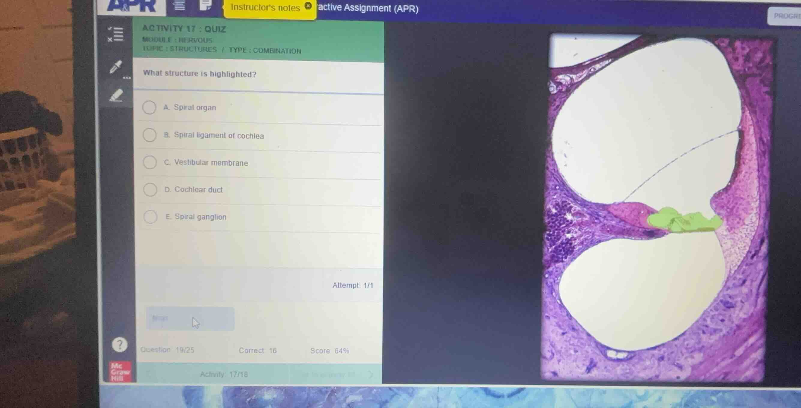
what structure is highlighted?
a. spiral organ
b. spiral ligament of cochlea
c. vestibular membrane
d. cochlear duct
e. spiral ganglion
Identify the histological section
The image displays a cross-section of a turn of the cochlea, showing the three fluid-filled chambers: the scala vestibuli (top), the cochlear duct (middle, scala media), and the scala tympani (bottom).
Analyze the highlighted structure
Using the Cochlea Histology knowledge point, we identify the specific region highlighted in green. This structure sits on the basilar membrane within the cochlear duct. It contains the sensory hair cells and supporting cells responsible for transducing sound vibrations into neural signals.
Correlate with anatomical terms
Using the Organ of Corti Anatomy knowledge point, we recognize that this specialized epithelial structure is known as the organ of Corti, or the spiral organ. The green highlight covers this entire sensory apparatus.
Evaluate the multiple-choice options
- A. Spiral organ: This matches the highlighted sensory structure on the basilar membrane.
- B. Spiral ligament of cochlea: This is the periosteal thickening on the outer wall (visible on the right side, colored pink).
- C. Vestibular membrane: This is the thin membrane separating the scala vestibuli from the cochlear duct (visible as the diagonal line).
- D. Cochlear duct: This refers to the entire middle triangular chamber, not just the specific highlighted sensory organ.
- E. Spiral ganglion: This is located within the modiolus (bony core) on the left side, containing the cell bodies of the bipolar sensory neurons.

Snap & solve any problem in the app
Get step-by-step solutions on Sovi AI
Photo-based solutions with guided steps
Explore more problems and detailed explanations
- (A) Spiral organ (Correct answer)
- (B) Spiral ligament of cochlea
- (C) Vestibular membrane
- (D) Cochlear duct
- (E) Spiral ganglion