QUESTION IMAGE
Question
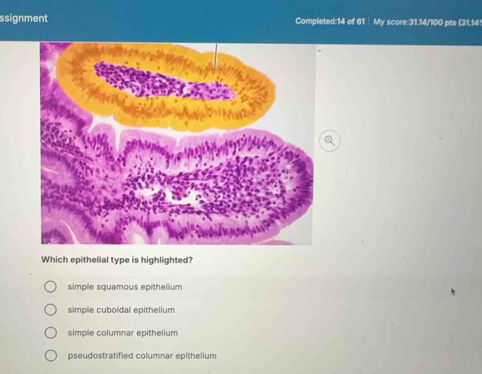
which epithelial type is highlighted?
simple squamous epithelium
simple cuboidal epithelium
simple columnar epithelium
pseudostratified columnar epithelium
Analyze the highlighted tissue
The image shows a histological section of the digestive tract (specifically, a cross-section of a villus and an adjacent fold). The highlighted region in yellow/orange outlines a single layer of tall, rectangular cells lining the lumen.
Identify cell shape and layers
Using Epithelial Tissue Types and Histological Identification, we observe that the nuclei are elongated and aligned in a neat row near the basal region of the cells. Since there is only one layer of these tall, column-like cells, this tissue is classified as a simple columnar epithelium.
Evaluate the options
- simple squamous epithelium: Composed of a single layer of flat, scale-like cells. This does not match the tall cells shown.
- simple cuboidal epithelium: Composed of a single layer of box-like, square cells with round, central nuclei.
- simple columnar epithelium: Composed of a single layer of tall, column-like cells with oval nuclei. This matches the highlighted tissue perfectly.
- pseudostratified columnar epithelium: Appears stratified because cell nuclei are at different levels, but all cells touch the basement membrane. The highlighted tissue shows a distinct, uniform single layer of nuclei.

Snap & solve any problem in the app
Get step-by-step solutions on Sovi AI
Photo-based solutions with guided steps
Explore more problems and detailed explanations
- (A) simple squamous epithelium
- (B) simple cuboidal epithelium
- (C) simple columnar epithelium (Correct answer)
- (D) pseudostratified columnar epithelium