QUESTION IMAGE
Question
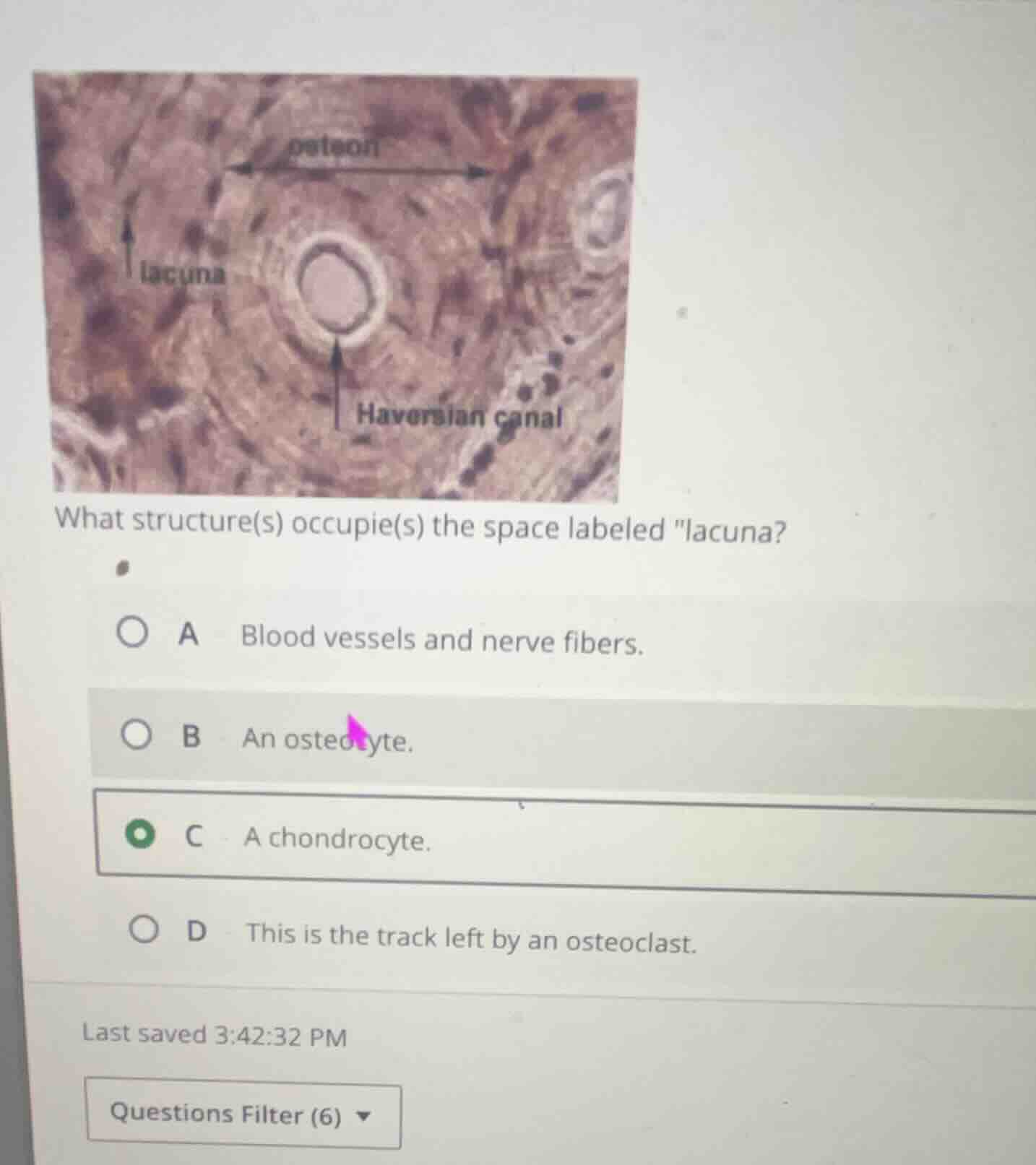
what structure(s) occupie(s) the space labeled \lacuna\?
a blood vessels and nerve fibers.
b an osteocyte.
c a chondrocyte.
d this is the track left by an osteoclast.
🆕 New Concept Discovered: Structure of Compact Bone
How bone tissue is organized at the microscopic level
Step 1: Identify the tissue type
The image shows a microscopic view of compact bone tissue. Key features of compact bone visible here include:
- Osteon: The basic structural unit of compact bone, consisting of concentric layers of bone matrix.
- Haversian canal: The central channel containing blood vessels and nerves.
- Lacunae: Small, dark, almond-shaped cavities arranged in concentric rings.
Step 2: Determine the contents of a lacuna
A lacuna (plural: lacunae) is a tiny space or cavity within the hard bone matrix.
- In living bone tissue, each lacuna is occupied by a mature bone cell called an osteocyte.
- Osteocytes maintain the surrounding bone matrix and communicate with neighboring cells through tiny channels called canaliculi.
Step 3: Evaluate the options
- A. Blood vessels and nerve fibers: These are located in the central Haversian canal, not the lacunae.
- B. An osteocyte: This is correct. Mature bone cells (osteocytes) reside inside the lacunae.
- C. A chondrocyte: Chondrocytes are cartilage cells. While they also live in lacunae, they are found in cartilage tissue, not bone tissue (osteons).
- D. This is the track left by an osteoclast: Osteoclasts are large cells that resorb (break down) bone tissue on bone surfaces; they do not live inside lacunae within osteons.

Snap & solve any problem in the app
Get step-by-step solutions on Sovi AI
Photo-based solutions with guided steps
Explore more problems and detailed explanations
B · An osteocyte.