QUESTION IMAGE
Question
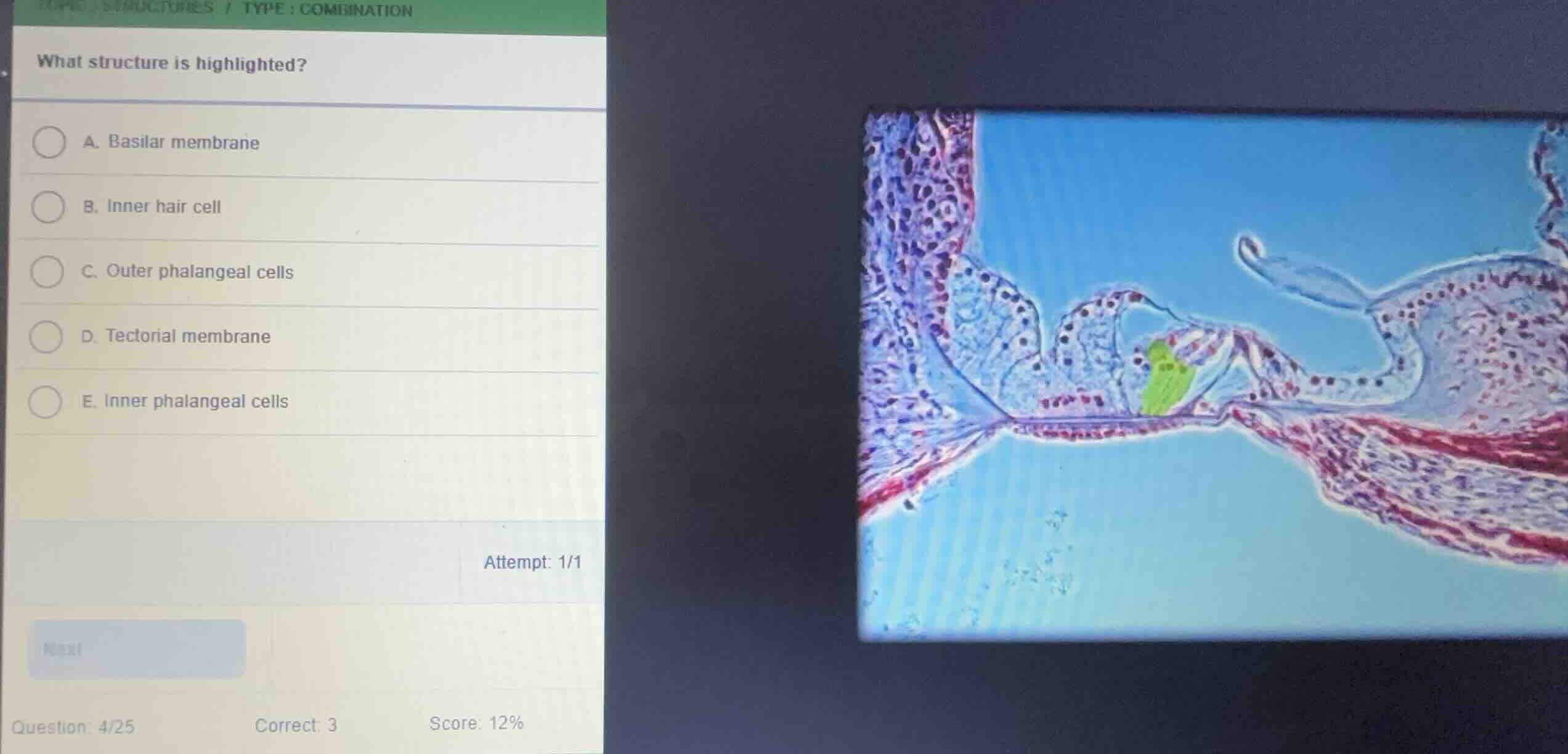
what structure is highlighted?
a. basilar membrane
b. inner hair cell
c. outer phalangeal cells
d. tectorial membrane
e. inner phalangeal cells
Identify the histological section
The image shows a cross-section of the cochlear duct, specifically focusing on the organ of Corti sitting on the basilar membrane.
Analyze the highlighted region
Using the Organ of Corti Anatomy knowledge point
The highlighted green region is located on the outer side of the tunnel of Corti, supporting the outer hair cells. These supporting cells are the outer phalangeal cells (also known as Deiters' cells), which support the basolateral aspects of the outer hair cells.
Match with options
- A. Basilar membrane: The fibrous layer beneath the organ of Corti.
- B. Inner hair cell: Located on the inner side of the tunnel of Corti, single row.
- C. Outer phalangeal cells: Supporting cells beneath and surrounding the outer hair cells (highlighted in green).
- D. Tectorial membrane: The acellular gelatinous structure projecting over the hair cells (visible above).
- E. Inner phalangeal cells: Supporting cells surrounding the inner hair cell on the inner side.
Therefore, the highlighted structure corresponds to the outer phalangeal cells.

Snap & solve any problem in the app
Get step-by-step solutions on Sovi AI
Photo-based solutions with guided steps
Explore more problems and detailed explanations
- (A) Basilar membrane
- (B) Inner hair cell
- (C) Outer phalangeal cells (Correct answer)
- (D) Tectorial membrane
- (E) Inner phalangeal cells