QUESTION IMAGE
Question
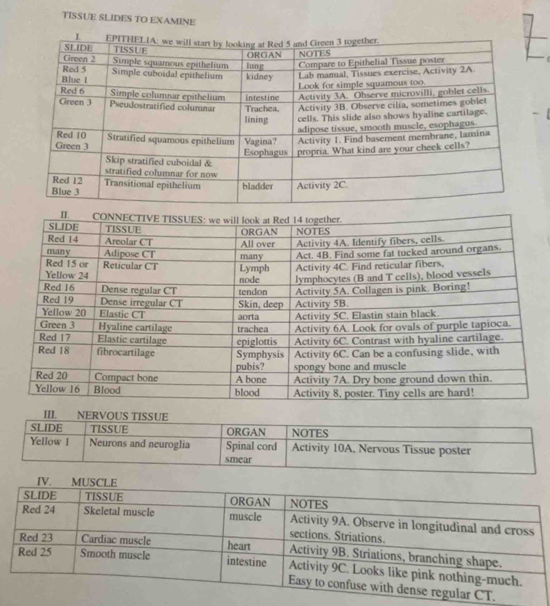
tissue slides to examine
i. epithelia: we will start by looking at red 5 and green 3 together.
\
ii. connective tissues: we will look at red 14 together.
\
iii. nervous tissue
\
iv. muscle
\
🆕 New Concept Discovered: Histological Tissue Classification
Identifying tissues by their structural types and organ locations.
Step 1: Analyze the Request
The user has provided an image of a lab reference sheet titled "TISSUE SLIDES TO EXAMINE". This sheet categorizes various tissue slides into four primary tissue types:
- Epithelia (Epithelial Tissues)
- Connective Tissues
- Nervous Tissue
- Muscle
To make this document highly accessible, searchable, and easy to study, we will transcribe the tables completely and accurately into clean Markdown tables.
Step 2: Transcribe Section I (Epithelia)
We extract the columns: SLIDE, TISSUE, ORGAN, and NOTES for the Epithelial tissue section.
| SLIDE | TISSUE | ORGAN | NOTES |
|---|---|---|---|
| Red 5 <br> Blue 1 | Simple cuboidal epithelium | kidney | Lab manual, Tissues exercise, Activity 2A. Look for simple squamous too. |
| Red 6 | Simple columnar epithelium | intestine | Activity 3A. Observe microvilli, goblet cells. |
| Green 3 | Pseudostratified columnar | Trachea, lining | Activity 3B. Observe cilia, sometimes goblet cells. This slide also shows hyaline cartilage, adipose tissue, smooth muscle, esophagus. |
| Red 10 <br> Green 3 | Stratified squamous epithelium | Vagina? <br> Esophagus | Activity 1. Find basement membrane, lamina propria. What kind are your cheek cells? |
| Skip stratified cuboidal & stratified columnar for now | |||
| Red 12 <br> Blue 3 | Transitional epithelium | bladder | Activity 2C. |
---
Step 3: Transcribe Section II (Connective Tissues)
We extract the details for the Connective Tissues section.
| SLIDE | TISSUE | ORGAN | NOTES |
|---|---|---|---|
| many | Adipose CT | many | Act. 4B. Find some fat tucked around organs. |
| Red 15 or <br> Yellow 24 | Reticular CT | Lymph node | Activity 4C. Find reticular fibers, lymphocytes (B and T cells), blood vessels |
| Red 16 | Dense regular CT | tendon | Activity 5A. Collagen is pink. Boring! |
| Red 19 | Dense irregular CT | Skin, deep | Activity 5B. |
| Yellow 20 | Elastic CT | aorta | Activity 5C. Elastin stain black. |
| Green 3 | Hyaline cartilage | trachea | Activity 6A. Look for ovals of purple tapioca. |
| Red 17 | Elastic cartilage | epiglottis | Activity 6C. Contrast with hyaline cartilage. |
| Red 18 | fibrocartilage | Symphysis pubis? | Activity 6C. Can be a confusing slide, with spongy bone and muscle |
| Red 20 | Compact bone | A bone | Activity 7A. Dry bone ground down thin. |
| Yellow 16 | Blood | blood | Activity 8, poster. Tiny cells are hard! |
---
Step 4: Transcribe Section III (Nervous Tissue)
We extract the details for the Nervous Tissue section.
| SLIDE | TISSUE | ORGAN | NOTES |
|---|
---
Step 5: Transcribe Section IV (Muscle)
We extract the details for the Muscle tissue section.
| SLIDE | TISSUE | ORGAN | NOTES |
|---|---|---|---|
| Red 23 | Cardiac muscle | heart | Activity 9B. Striations, branching shape. |
| Red 25 | Smooth muscle | intestine | Activity 9C. Looks like pink nothing-much. Easy to confuse with dense regular CT. |

Snap & solve any problem in the app
Get step-by-step solutions on Sovi AI
Photo-based solutions with guided steps
Explore more problems and detailed explanations
Here is the complete transcription of your Tissue Slides to Examine lab sheet:
I. EPITHELIA (starting with Red 5 and Green 3 together)
| SLIDE | TISSUE | ORGAN | NOTES |
|---|---|---|---|
| Red 5 <br> Blue 1 | Simple cuboidal epithelium | kidney | Lab manual, Tissues exercise, Activity 2A. Look for simple squamous too. |
| Red 6 | Simple columnar epithelium | intestine | Activity 3A. Observe microvilli, goblet cells. |
| Green 3 | Pseudostratified columnar | Trachea, lining | Activity 3B. Observe cilia, sometimes goblet cells. This slide also shows hyaline cartilage, adipose tissue, smooth muscle, esophagus. |
| Red 10 <br> Green 3 | Stratified squamous epithelium | Vagina? <br> Esophagus | Activity 1. Find basement membrane, lamina propria. What kind are your cheek cells? |
| Skip stratified cuboidal & stratified columnar for now | |||
| Red 12 <br> Blue 3 | Transitional epithelium | bladder | Activity 2C. |
---
II. CONNECTIVE TISSUES (starting with Red 14 together)
| SLIDE | TISSUE | ORGAN | NOTES |
|---|---|---|---|
| many | Adipose CT | many | Act. 4B. Find some fat tucked around organs. |
| Red 15 or <br> Yellow 24 | Reticular CT | Lymph node | Activity 4C. Find reticular fibers, lymphocytes (B and T cells), blood vessels |
| Red 16 | Dense regular CT | tendon | Activity 5A. Collagen is pink. Boring! |
| Red 19 | Dense irregular CT | Skin, deep | Activity 5B. |
| Yellow 20 | Elastic CT | aorta | Activity 5C. Elastin stain black. |
| Green 3 | Hyaline cartilage | trachea | Activity 6A. Look for ovals of purple tapioca. |
| Red 17 | Elastic cartilage | epiglottis | Activity 6C. Contrast with hyaline cartilage. |
| Red 18 | fibrocartilage | Symphysis pubis? | Activity 6C. Can be a confusing slide, with spongy bone and muscle |
| Red 20 | Compact bone | A bone | Activity 7A. Dry bone ground down thin. |
| Yellow 16 | Blood | blood | Activity 8, poster. Tiny cells are hard! |
---
III. NERVOUS TISSUE
| SLIDE | TISSUE | ORGAN | NOTES |
|---|
---
IV. MUSCLE
| SLIDE | TISSUE | ORGAN | NOTES |
|---|---|---|---|
| Red 23 | Cardiac muscle | heart | Activity 9B. Striations, branching shape. |
| Red 25 | Smooth muscle | intestine | Activity 9C. Looks like pink nothing-much. Easy to confuse with dense regular CT. |