QUESTION IMAGE
Question
review sheet
tissues
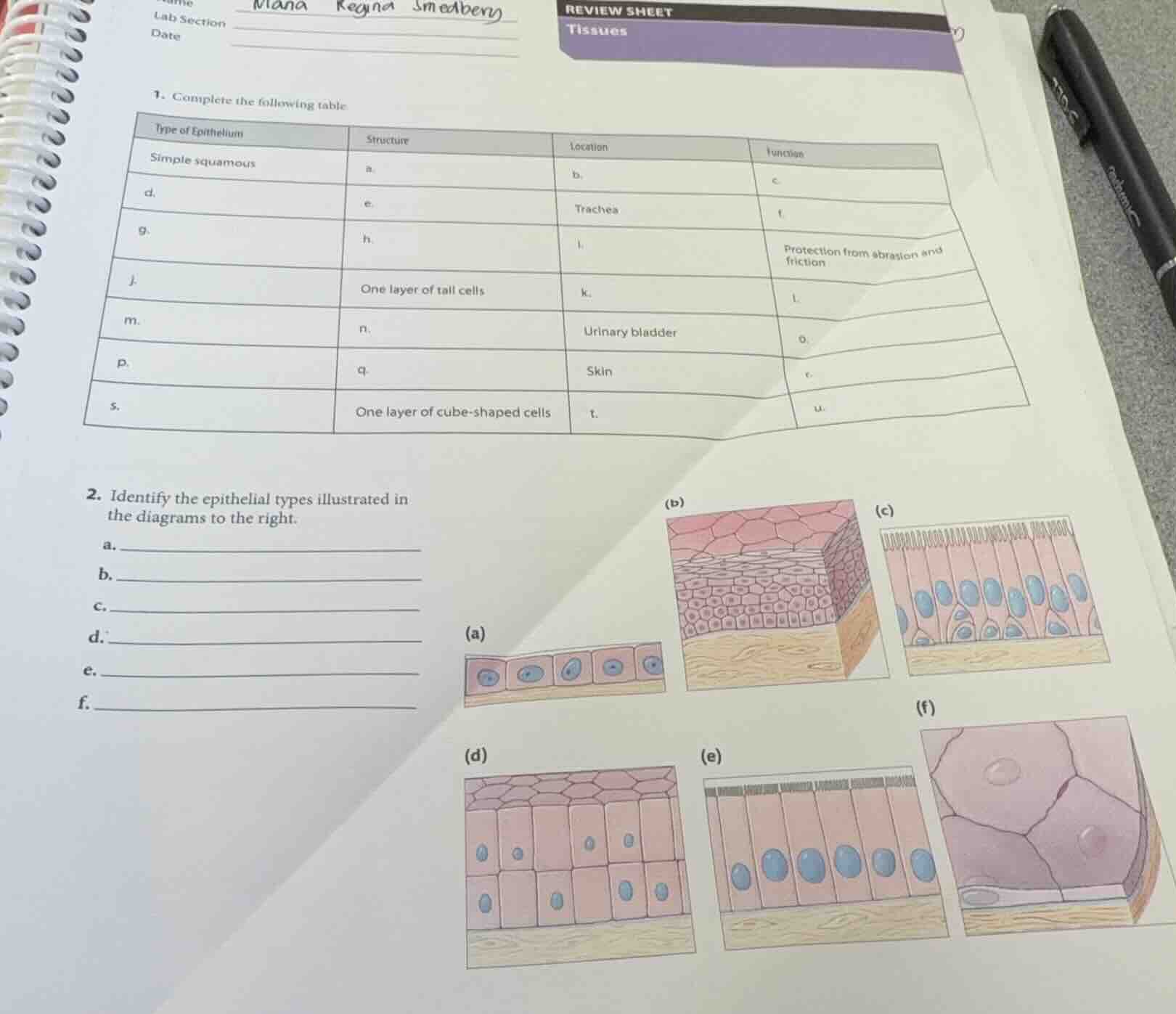
- complete the following table:
\
$$\begin{tabular}{|l|l|l|l|}
\\hline
type of epithelium & structure & location & function \\\\
\\hline
simple squamous & a. & b. & c. \\\\
\\hline
d. & e. & trachea & f. \\\\
\\hline
g. & h. & i. & protection from abrasion and friction \\\\
\\hline
j. & one layer of tall cells & k. & l. \\\\
\\hline
m. & n. & urinary bladder & o. \\\\
\\hline
p. & q. & skin & r. \\\\
\\hline
s. & one layer of cube-shaped cells & t. & u. \\\\
\\hline
\\end{tabular}$$
- identify the epithelial types illustrated in the diagrams to the right.
a.
b.
c.
d.
e.
f.
🆕 New Concept Discovered: Epithelial Tissue Classification
How structure dictates location and function in tissues.
Step 1: Analyze Simple Squamous Epithelium (Row 1)
- Structure (a): Simple squamous epithelium consists of a single layer of flat, scale-like cells.
- Location (b): Commonly found where rapid diffusion or filtration is required, such as the air sacs of the lungs (alveoli) or the lining of blood vessels (endothelium).
- Function (c): Facilitates diffusion, filtration, and secretion of lubricating substances.
Step 2: Analyze Trachea Epithelium (Row 2)
- Type of Epithelium (d): The trachea is lined by pseudostratified ciliated columnar epithelium.
- Structure (e): Appears stratified because cell nuclei lie at different levels, but it is a single layer of cells of varying heights, typically ciliated.
- Function (f): Secretes mucus (via goblet cells) and propels it upward using cilia to protect the respiratory tract.
Step 3: Analyze Protection from Abrasion (Row 3)
- Function: Protection from abrasion and friction.
- Type of Epithelium (g): Stratified squamous epithelium (non-keratinized, as it is typically found in moist internal areas subject to wear and tear).
- Structure (h): Multiple layers of cells; basal cells are cuboidal/columnar, while apical surface cells are flattened (squamous).
- Location (i): Lining of the esophagus, mouth, and vagina.
Step 4: Analyze One Layer of Tall Cells (Row 4)
- Structure: One layer of tall cells.
- Type of Epithelium (j): Simple columnar epithelium.
- Location (k): Lining of the digestive tract (stomach to anal canal) or gallbladder.
- Function (l): Absorption of nutrients and secretion of mucus and enzymes.
Step 5: Analyze Urinary Bladder Epithelium (Row 5)
- Location: Urinary bladder.
- Type of Epithelium (m): Transitional epithelium.
- Structure (n): Stratified layers of cells; surface cells are dome-shaped (umbrella cells) when relaxed, but flatten out when stretched.
- Function (o): Stretches readily and permits distension of the urinary organ as it fills with urine.
Step 6: Analyze Skin Epithelium (Row 6)
- Location: Skin (epidermis).
- Type of Epithelium (p): Keratinized stratified squamous epithelium.
- Structure (q): Multiple layers of cells; surface cells are dead, flat, and filled with the tough protein keratin.
- Function (r): Protects underlying tissues from mechanical damage, pathogens, and dehydration.
Step 7: Analyze One Layer of Cube-Shaped Cells (Row 7)
- Structure: One layer of cube-shaped cells.
- Type of Epithelium (s): Simple cuboidal epithelium.
- Location (t): Kidney tubules, ducts of small glands, and ovary surface.
- Function (u): Secretion and absorption.
Step 8: Identify Diagrams in Question 2
- Diagram (a): A single layer of cube-shaped cells with central, spherical nuclei. This is simple cuboidal epithelium.
- Diagram (b): Multiple layers of cells, with flat, scale-like cells at the apical surface. This is stratified squamous epithelium.
- Diagram (c): A single layer of cells of differing heights with nuclei at different levels, bearing cilia at the apical surface. This is pseudostratified ciliated columnar epithelium.
- Diagram (d): Multiple layers of cells where the surface cells are rounded/dome-shaped. This is transitional epithelium.
- Diagram (e): A single layer of tall, column-like cells with oval nuclei located near the base. This is simple columnar epithelium.
- **Diagram (f…

Snap & solve any problem in the app
Get step-by-step solutions on Sovi AI
Photo-based solutions with guided steps
Explore more problems and detailed explanations
1. Complete the following table:
| Letter | Answer |
|---|---|
| b. | Air sacs of lungs (alveoli) / Lining of blood vessels |
| c. | Diffusion, filtration, and secretion |
| d. | Pseudostratified ciliated columnar |
| e. | Single layer of cells of differing heights, nuclei at different levels, ciliated |
| f. | Secretion and propulsion of mucus |
| g. | Stratified squamous (non-keratinized) |
| h. | Multiple layers of cells; apical cells are flat |
| i. | Lining of the esophagus, mouth, and vagina |
| j. | Simple columnar |
| k. | Lining of the digestive tract (stomach to rectum) |
| l. | Absorption and secretion of mucus and enzymes |
| m. | Transitional |
| n. | Stratified; surface cells are dome-shaped/rounded when relaxed |
| o. | Stretches and permits distension of the bladder |
| p. | Stratified squamous (keratinized) |
| q. | Multiple layers of cells; apical cells are dead, flat, and keratinized |
| r. | Protection against abrasion, pathogens, and water loss |
| s. | Simple cuboidal |
| t. | Kidney tubules / Ducts of small glands |
| u. | Secretion and absorption |
---
2. Identify the epithelial types illustrated in the diagrams:
- a. Simple cuboidal epithelium
- b. Stratified squamous epithelium
- c. Pseudostratified ciliated columnar epithelium
- d. Transitional epithelium
- e. Simple columnar epithelium
- f. Simple squamous epithelium