QUESTION IMAGE
Question
pre-lab video coaching activity: examining a long bone
5 parts | 7 of 15 points
osteon
lamella
trabeculae
perforating fibers
periosteum
epiphyseal plate
endosteum
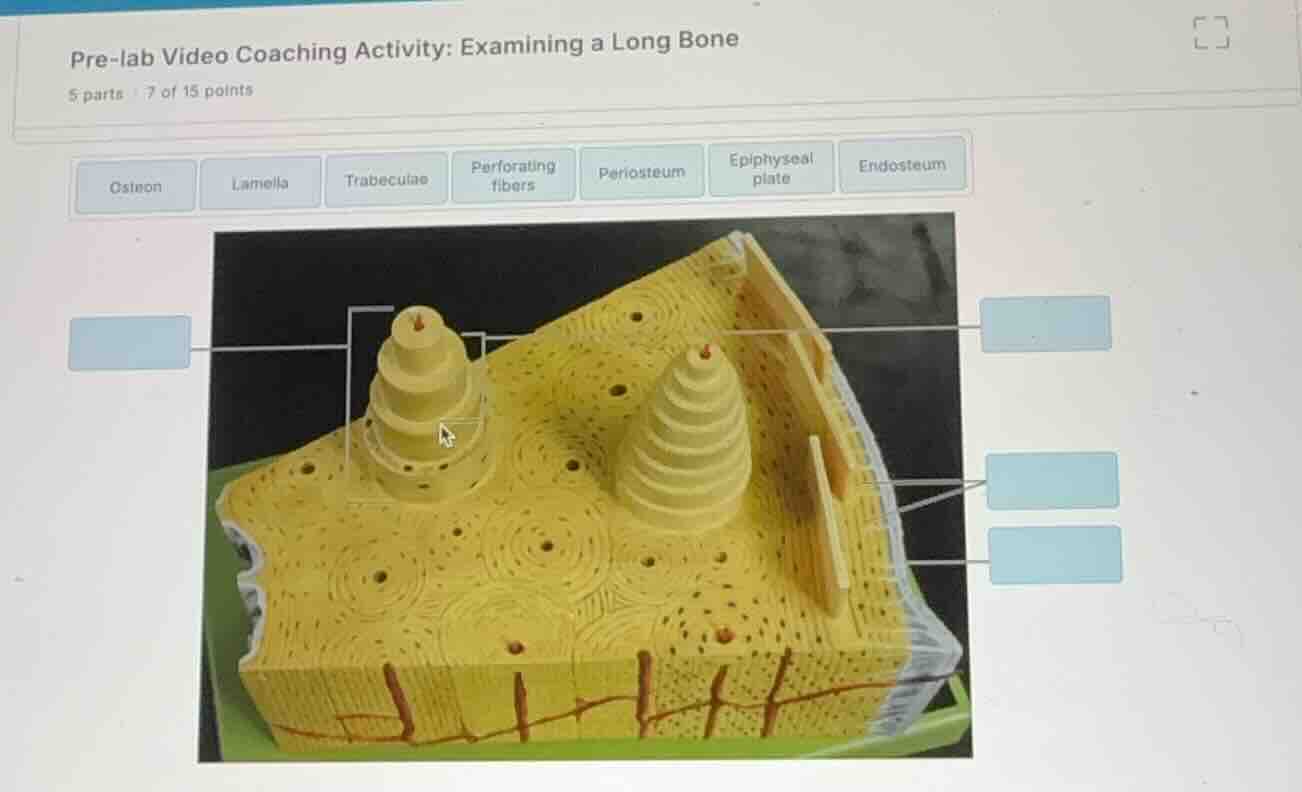
Identify the anatomical model
The image shows a 3D anatomical model of compact bone tissue histology, highlighting the structural units of bone.
Analyze the left labels
Using the Compact Bone Anatomy knowledge point
- The bracket on the far left points to an entire structural cylinder unit of compact bone. This unit is the Osteon.
- The line branching from that bracket points to an individual concentric ring layer within the osteon. This layer is the Lamella.
Analyze the right labels
Using the Compact Bone Anatomy knowledge point
- The top-right label points to the inner membrane lining the bony canals and covering trabeculae. In this model, it points to the inner lining of the central canal or inner surface, which is the Endosteum.
- The middle-right label points to the tough outer fibrous membrane covering the bone surface, which is the Periosteum.
- The bottom-right label points to the collagen fibers anchoring this outer membrane to the underlying bone matrix, which are the Perforating fibers (Sharpey's fibers).
Verify unused options
- Trabeculae and Epiphyseal plate are not shown in this compact bone wedge model (trabeculae are found in spongy bone, and the epiphyseal plate is located at the metaphysis of growing long bones).

Snap & solve any problem in the app
Get step-by-step solutions on Sovi AI
Photo-based solutions with guided steps
Explore more problems and detailed explanations
| Label Position | Anatomical Structure |
|---|---|
| Left (line pointing to concentric layer) | Lamella |
| Top Right | Endosteum |
| Middle Right | Periosteum |
| Bottom Right | Perforating fibers |