QUESTION IMAGE
Question
place the appropriate labels in their respective targets.
show instructions
columnar cell, simple epithelium, stratified epithelium, squamous cell, cuboidal cell
Identify tissue layers
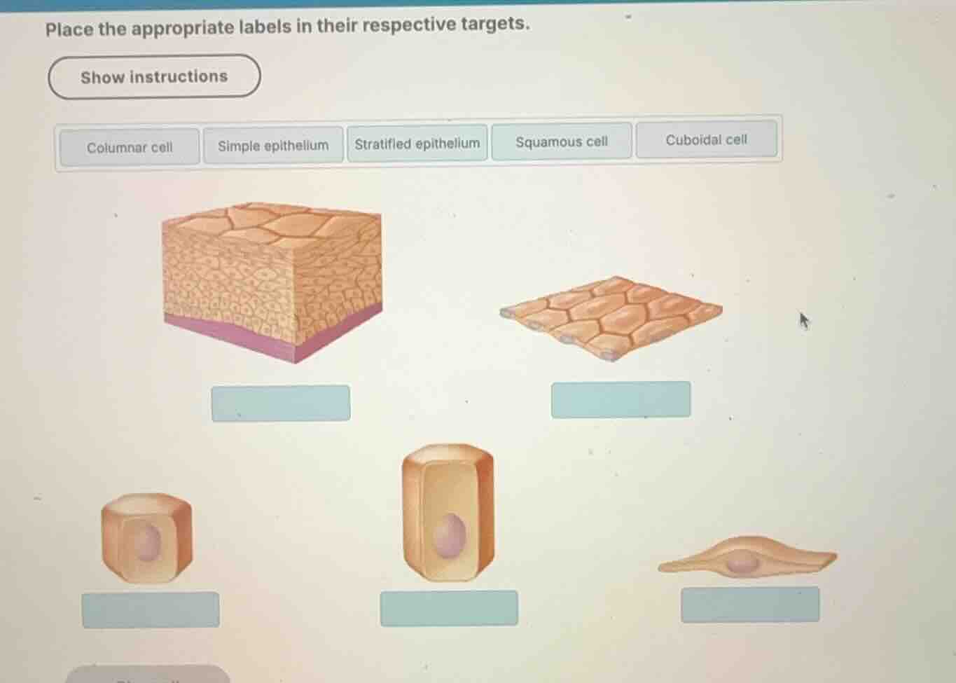
Using the Epithelial Tissue Classification knowledge point, we classify epithelial tissues by the number of cell layers. The top-left diagram shows multiple layers of cells stacked on top of each other, which represents a stratified epithelium. The top-right diagram shows a single layer of flat cells, which represents a simple epithelium.
Identify cell shapes
We classify individual epithelial cells by their physical shape:
- The bottom-left cell is cube-shaped with a central, round nucleus, representing a cuboidal cell.
- The bottom-middle cell is tall and column-like with an elongated nucleus, representing a columnar cell.
- The bottom-right cell is flat and scale-like with a flattened nucleus, representing a squamous cell.
Match labels to targets
We align each of the five targets with the correct label from the top options:
- Top-left diagram (multi-layered tissue): "Stratified epithelium"
- Top-right diagram (single-layered tissue): "Simple epithelium"
- Bottom-left cell (cube-shaped): "Cuboidal cell"
- Bottom-middle cell (tall/column-shaped): "Columnar cell"
- Bottom-right cell (flat/scale-shaped): "Squamous cell"

Snap & solve any problem in the app
Get step-by-step solutions on Sovi AI
Photo-based solutions with guided steps
Explore more problems and detailed explanations
- Top-left target (multi-layered tissue): Stratified epithelium
- Top-right target (single-layered tissue): Simple epithelium
- Bottom-left target (cube-shaped cell): Cuboidal cell
- Bottom-middle target (tall cell): Columnar cell
- Bottom-right target (flat cell): Squamous cell