QUESTION IMAGE
Question
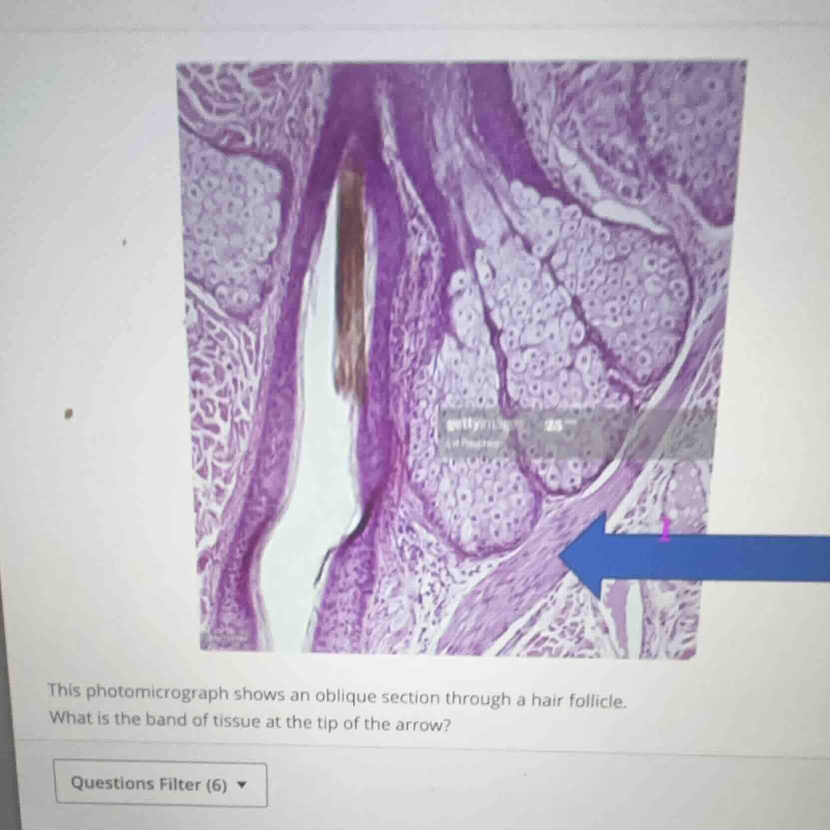
this photomicrograph shows an oblique section through a hair follicle.
what is the band of tissue at the tip of the arrow?
🆕 New Concept Discovered: Integumentary Histology Identification
Identifying microscopic structures of the skin and hair follicle.
Step 1: Analyze the image and anatomical context
The photomicrograph shows a section through a hair follicle in the dermis of the skin.
- In the center, we see a hair follicle containing a hair shaft.
- Adjacent to the follicle are large, pale-staining, multi-lobular glands, which are sebaceous glands.
- The blue arrow points to a distinct, elongated band of smooth muscle tissue extending at an angle from the connective tissue sheath of the hair follicle toward the upper dermis.
Step 2: Identify the specific tissue band
The band of smooth muscle associated with a hair follicle is the arrector pili muscle.
- Function: When this muscle contracts, it pulls the hair follicle upright, causing the hair to stand on end (commonly known as "goosebumps") and helping to squeeze sebum out of the sebaceous gland into the hair follicle.
- Histological appearance: It consists of a bundle of smooth muscle fibers, which appear as elongated, eosinophilic (pinkish/purple) cells with centrally located, elongated nuclei.

Snap & solve any problem in the app
Get step-by-step solutions on Sovi AI
Photo-based solutions with guided steps
Explore more problems and detailed explanations
Arrector pili muscle