QUESTION IMAGE
Question
module: nervous
topic: structures / type: combination
what structure is highlighted?
a. outer phalangeal cells
b. outer hair cell
c. inner phalangeal cells
d. tectorial membrane
e. inner hair cell
Identify the histological section
Using the Organ of Corti Anatomy knowledge point
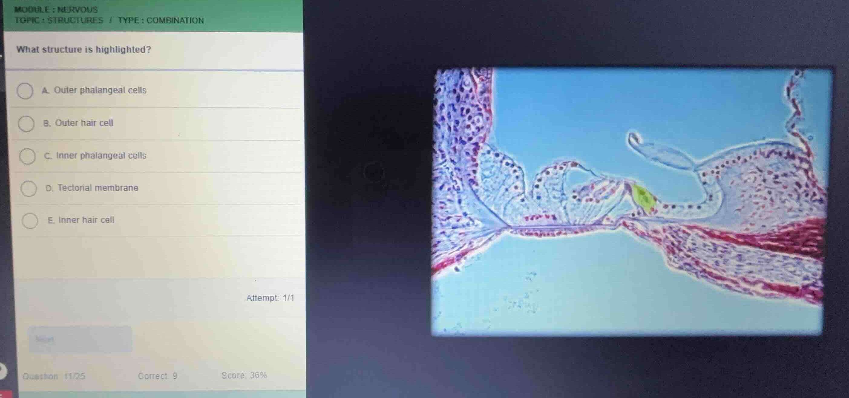
The provided image shows a high-magnification histological section of the organ of Corti within the cochlea of the inner ear, resting on the basilar membrane.
Locate the highlighted structure
Using the Inner Hair Cell Histology and Inner Phalangeal Cells knowledge points
The highlighted green region is located on the inner (medial) side of the inner tunnel of Corti. It represents a single, flask-shaped sensory cell situated on the inner side of the inner pillar cell, supported by an inner phalangeal cell. This specific cell is the single row of inner hair cells.
Differentiate from other options
Using the Organ of Corti Anatomy knowledge point
- Outer hair cells and outer phalangeal cells are located on the opposite (lateral) side of the inner tunnel of Corti, arranged in three to four rows.
- The tectorial membrane is the acellular gel-like structure projecting over the hair cells, which is visible but not highlighted in green.
- The highlighted cell is the flask-shaped sensory cell itself, which is the inner hair cell.

Snap & solve any problem in the app
Get step-by-step solutions on Sovi AI
Photo-based solutions with guided steps
Explore more problems and detailed explanations
- (A) Outer phalangeal cells
- (B) Outer hair cell
- (C) Inner phalangeal cells
- (D) Tectorial membrane
- (E) Inner hair cell (Correct answer)