QUESTION IMAGE
Question
dura mater
synovial fluid
subarachnoid space
epidural space
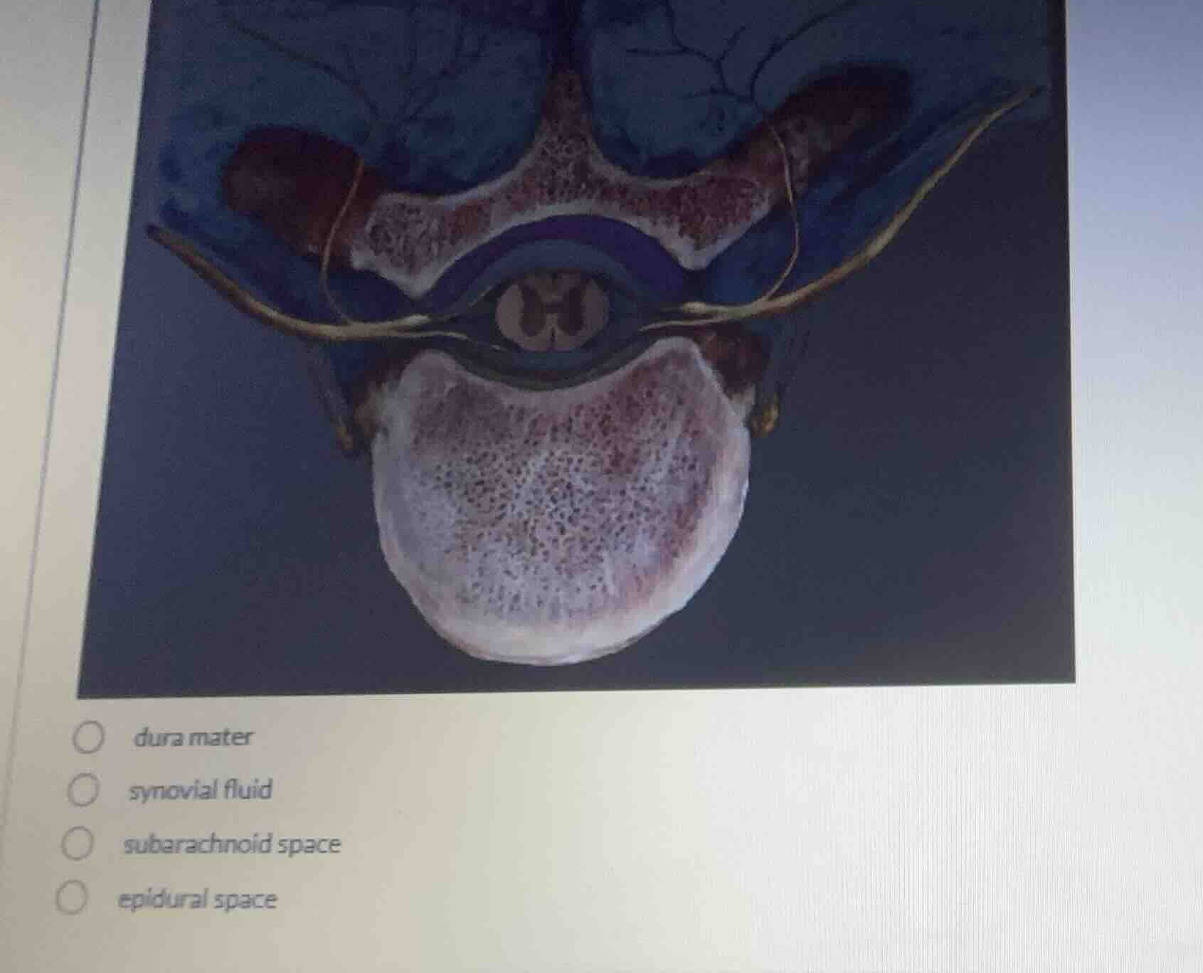
Identify the anatomical structure highlighted in blue
The image shows a cross-section of a vertebra containing the spinal cord and its surrounding meningeal layers. The region highlighted in bright blue is the space located outside the dura mater, between the dural sheath and the bony wall of the vertebral canal. Using the Epidural Space Anatomy and Spinal Meninges knowledge points, we identify this outermost space as the epidural space.
Verify the anatomical boundaries
The spinal meninges consist of three layers: the innermost pia mater, the middle arachnoid mater, and the outermost dura mater. The space between the arachnoid mater and pia mater is the subarachnoid space, which contains cerebrospinal fluid. The space outside the dura mater, within the vertebral canal, is the epidural space, which contains adipose tissue and blood vessels. The blue highlighted region clearly represents this outermost protective cushion.

Snap & solve any problem in the app
Get step-by-step solutions on Sovi AI
Photo-based solutions with guided steps
Explore more problems and detailed explanations
- dura mater
- synovial fluid
- subarachnoid space
- epidural space (Correct answer)