QUESTION IMAGE
Question
complete the labeling activity.
place the appropriate labels in their respective targets.
show instructions
dermal papillae
stratum basale
hypodermis
sweat pore
sweat gland
epidermis
sensory neurons
dermis
stratum corneum
stratum spinosum
<pre_analysis>
{
"quality": "clear",
"question_count": 1,
"skills_matched": [
"direct_cot",
"explain_then_answer",
"tutor"
],
"needs_drawing": false,
"concepts_used": [],
"new_concepts": [
"Integumentary System Anatomy",
"Skin Layers",
"Epidermal Strata"
],
"current_concepts": [
"Integumentary System Anatomy",
"Skin Layers",
"Epidermal Strata"
]
}
</pre_analysis>
<reasoning>
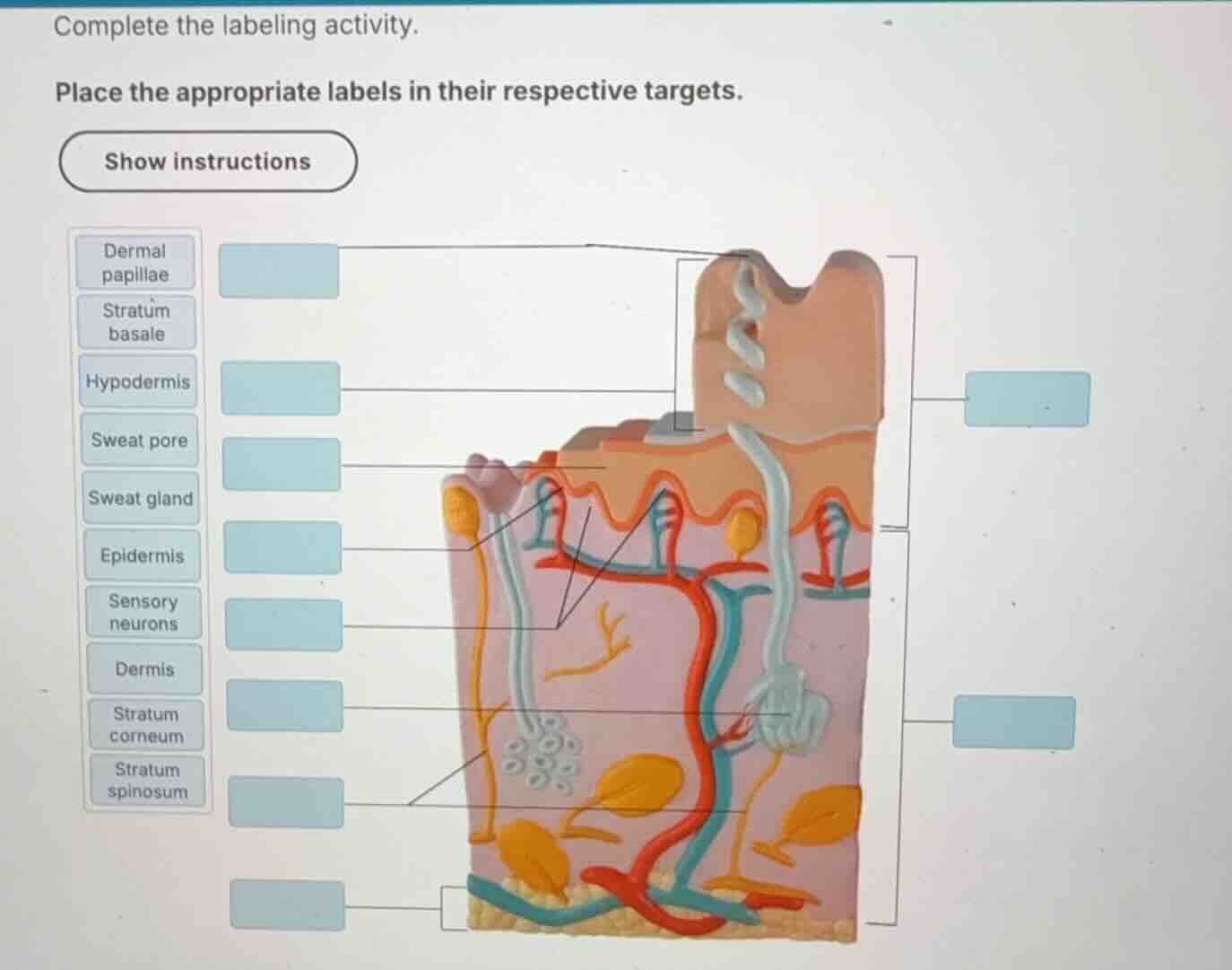
Identify epidermal layers and structures
The diagram shows a 3D cross-section of human skin. Let's trace the pointers on the left side from top to bottom:
- The first pointer at the very top left points to the opening of the sweat duct on the skin surface: Sweat pore.
- The second pointer points to the outermost, thick keratinized layer of the epidermis: Stratum corneum.
- The third pointer points to the middle layer of the epidermis: Stratum spinosum.
- The fourth pointer points to the deepest, single-cell layer of the epidermis: Stratum basale.
- The fifth pointer points to the finger-like projections of the dermis extending into the epidermis: Dermal papillae.
- The sixth pointer points to the yellow nerve fibers in the dermis: Sensory neurons.
- The seventh pointer points to the coiled tubular gland in the dermis: Sweat gland.
- The eighth pointer at the bottom left points to the subcutaneous layer containing adipose tissue: Hypodermis.
Identify major skin regions
Now let's trace the brackets on the right side:
- The upper bracket spans the entire outer epithelial region: Epidermis.
- The lower bracket spans the thick connective tissue region beneath the epidermis: Dermis.
</reasoning>
<answer>
The correct labels for the targets in the diagram are:
Left Column (Top to Bottom):
- Sweat pore
- Stratum corneum
- Stratum spinosum
- Stratum basale
- Dermal papillae
- Sensory neurons
- Sweat gland
- Hypodermis
Right Column Brackets (Top to Bottom):
- Epidermis (upper bracket)
- Dermis (lower bracket)
</answer>
<post_analysis>
{
"subject": "Natural Science",
"question_type": "Fill-in-the-blank",
"knowledge_point": [
"Natural Science",
"Anatomy & Physiology",
"Integumentary System"
]
}
</post_analysis>

Snap & solve any problem in the app
Get step-by-step solutions on Sovi AI
Photo-based solutions with guided steps
Explore more problems and detailed explanations
<pre_analysis>
{
"quality": "clear",
"question_count": 1,
"skills_matched": [
"direct_cot",
"explain_then_answer",
"tutor"
],
"needs_drawing": false,
"concepts_used": [],
"new_concepts": [
"Integumentary System Anatomy",
"Skin Layers",
"Epidermal Strata"
],
"current_concepts": [
"Integumentary System Anatomy",
"Skin Layers",
"Epidermal Strata"
]
}
</pre_analysis>
<reasoning>
Identify epidermal layers and structures
The diagram shows a 3D cross-section of human skin. Let's trace the pointers on the left side from top to bottom:
- The first pointer at the very top left points to the opening of the sweat duct on the skin surface: Sweat pore.
- The second pointer points to the outermost, thick keratinized layer of the epidermis: Stratum corneum.
- The third pointer points to the middle layer of the epidermis: Stratum spinosum.
- The fourth pointer points to the deepest, single-cell layer of the epidermis: Stratum basale.
- The fifth pointer points to the finger-like projections of the dermis extending into the epidermis: Dermal papillae.
- The sixth pointer points to the yellow nerve fibers in the dermis: Sensory neurons.
- The seventh pointer points to the coiled tubular gland in the dermis: Sweat gland.
- The eighth pointer at the bottom left points to the subcutaneous layer containing adipose tissue: Hypodermis.
Identify major skin regions
Now let's trace the brackets on the right side:
- The upper bracket spans the entire outer epithelial region: Epidermis.
- The lower bracket spans the thick connective tissue region beneath the epidermis: Dermis.
</reasoning>
<answer>
The correct labels for the targets in the diagram are:
Left Column (Top to Bottom):
- Sweat pore
- Stratum corneum
- Stratum spinosum
- Stratum basale
- Dermal papillae
- Sensory neurons
- Sweat gland
- Hypodermis
Right Column Brackets (Top to Bottom):
- Epidermis (upper bracket)
- Dermis (lower bracket)
</answer>
<post_analysis>
{
"subject": "Natural Science",
"question_type": "Fill-in-the-blank",
"knowledge_point": [
"Natural Science",
"Anatomy & Physiology",
"Integumentary System"
]
}
</post_analysis>