QUESTION IMAGE
Question
activity 17: quiz
module: nervous
topic: structures / type: combination
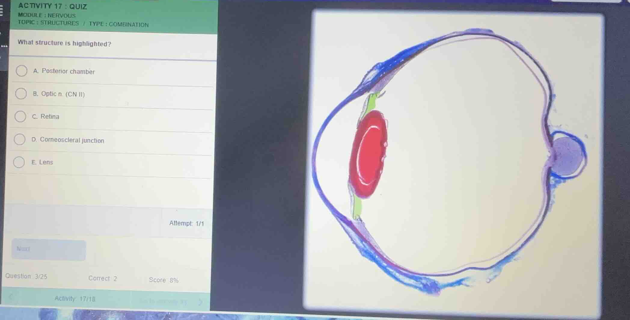
what structure is highlighted?
a. posterior chamber
b. optic n. (cn ii)
c. retina
d. corneoscleral junction
e. lens
Identify the highlighted structure
Using the Eye Anatomy knowledge point
The provided histological cross-section of the eye shows several structures highlighted in different colors:
- The outer fibrous layer (cornea and sclera) is highlighted in blue.
- The ciliary body/iris region is highlighted in green.
- The structure highlighted in bright red is the biconvex, transparent structure situated behind the iris, which is the lens.
Evaluate the given options
- A. Posterior chamber: This is the narrow space filled with aqueous humor behind the iris and in front of the lens (not highlighted in red).
- B. Optic n. (CN II): This is the nerve exiting the posterior aspect of the eye (highlighted in purple/blue on the right).
- C. Retina: This is the inner neural layer lining the back of the eye (not highlighted in red).
- D. Corneoscleral junction: This is the transition zone between the cornea and sclera (not highlighted in red).
- E. Lens: This matches the biconvex structure highlighted in bright red in the anterior segment of the eye.

Snap & solve any problem in the app
Get step-by-step solutions on Sovi AI
Photo-based solutions with guided steps
Explore more problems and detailed explanations
- (A) Posterior chamber
- (B) Optic n. (CN II)
- (C) Retina
- (D) Corneoscleral junction
- (E) Lens (Correct answer)