QUESTION IMAGE
Question
activity 17 : quiz
module : nervous
topic : structures / type : combination
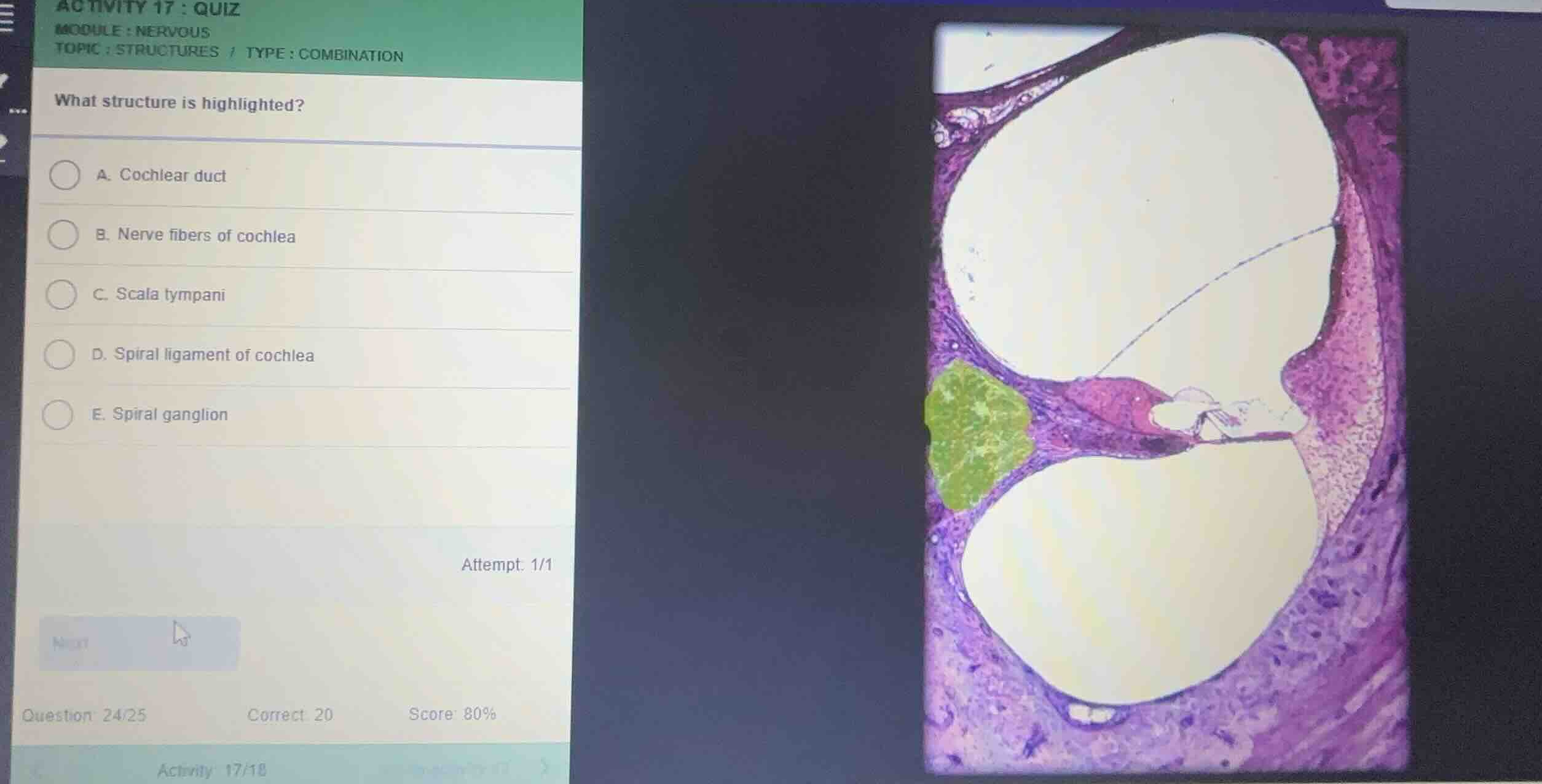
what structure is highlighted?
a. cochlear duct
b. nerve fibers of cochlea
c. scala tympani
d. spiral ligament of cochlea
e. spiral ganglion
attempt: 1/1
question: 24/25
correct: 20
score: 80%
Identify the highlighted region
Using the Cochlea Histology knowledge point, we examine the cross-section of the cochlear turn. The image shows the three fluid-filled chambers: the scala vestibuli (top), the cochlear duct (middle), and the scala tympani (bottom). The modiolus is located on the left side of this section.
Determine the specific structure
Using the Spiral Ganglion Histology knowledge point
The green highlighted cluster of neuronal cell bodies is located within the bony modiolus (specifically within Rosenthal's canal). These bipolar neurons form the spiral ganglion, which gives rise to the auditory nerve fibers.

Snap & solve any problem in the app
Get step-by-step solutions on Sovi AI
Photo-based solutions with guided steps
Explore more problems and detailed explanations
- (A) Cochlear duct
- (B) Nerve fibers of cochlea
- (C) Scala tympani
- (D) Spiral ligament of cochlea
- (E) Spiral ganglion (Correct answer)