QUESTION IMAGE
Question
activity 17: quiz
module: nervous
topic: structures / type: combination
what structure is highlighted?
a. corneoscleral junction
b. retina
c. sclera
d. ciliary body
e. anterior cavity
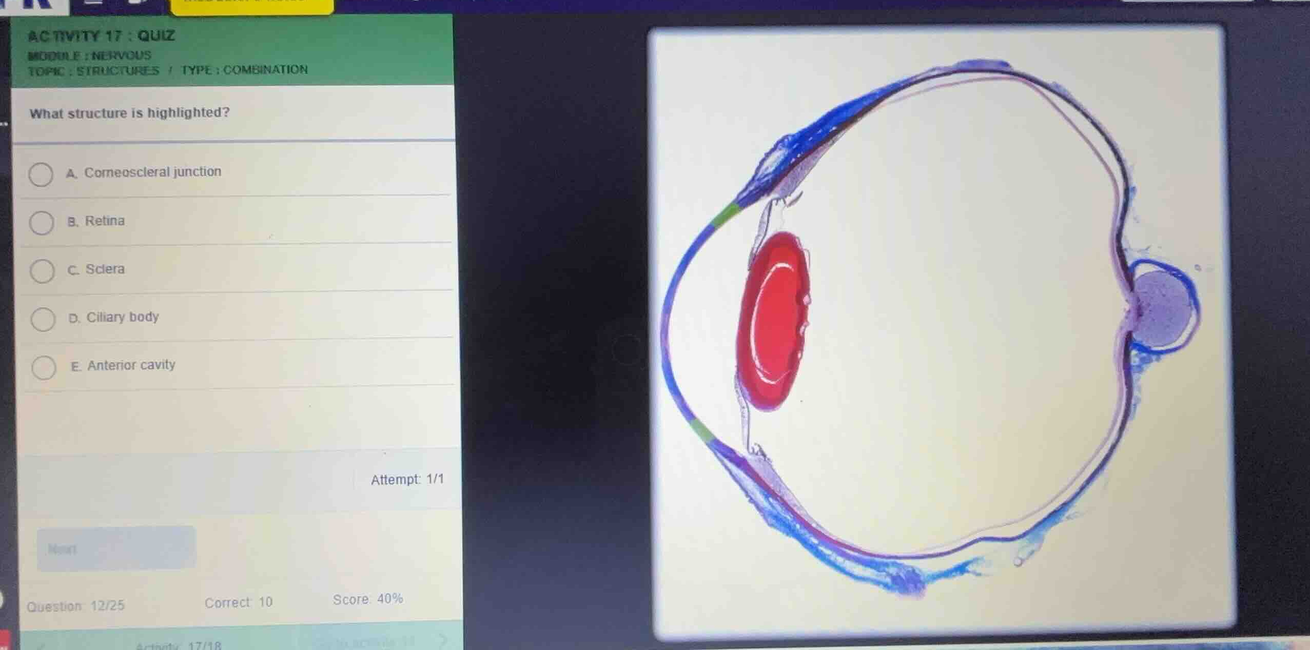
Identify the highlighted region
Using the Eye Anatomy and Ocular Histology knowledge points, we analyze the histological cross-section of the eye. The highlighted region is colored green on the anterior portion of the fibrous tunic, specifically at the transition zone where the transparent cornea meets the opaque sclera.
Analyze the anatomical transition
Using the Ocular Histology knowledge point, this specific boundary region where the cornea transitions into the sclera is anatomically defined as the limbus or corneoscleral junction.
Match with the options
Using the Eye Anatomy knowledge point, we compare the highlighted green region with the provided choices:
- A. Corneoscleral junction: This matches the transition zone highlighted in green.
- B. Retina: The inner neural layer, shown in pink/purple along the posterior wall, which is not highlighted in green.
- C. Sclera: The outer fibrous layer, shown in blue, which is not highlighted in green.
- D. Ciliary body: The thickened vascular tunic region posterior to the iris, which is not highlighted in green.
- E. Anterior cavity: The fluid-filled space anterior to the lens, which is not highlighted.
Therefore, the highlighted structure is the corneoscleral junction.

Snap & solve any problem in the app
Get step-by-step solutions on Sovi AI
Photo-based solutions with guided steps
Explore more problems and detailed explanations
- (A) Corneoscleral junction (Correct answer)
- (B) Retina
- (C) Sclera
- (D) Ciliary body
- (E) Anterior cavity