QUESTION IMAGE
Question
activity 10: quiz
module: cardiovascular
topic: structures / type: multiple choice (mc)
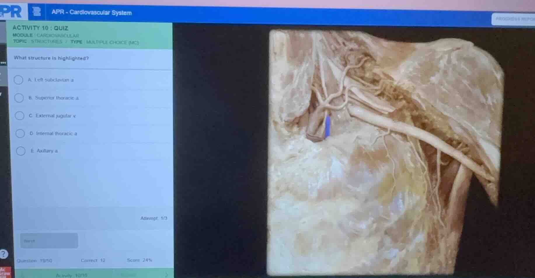
what structure is highlighted?
a. left subclavian a.
b. superior thoracic a.
c. external jugular v.
d. internal thoracic a.
e. axillary a.
Identify the anatomical region
The image shows a cadaveric dissection of the anterior thoracic wall, axilla, and shoulder region. The clavicle has been cut or reflected to expose the deeper neurovascular structures traveling from the root of the neck into the upper limb.
Analyze the highlighted structure
A blue highlight is placed on a major vessel located deep to the clavicular region, continuing directly into the arm. This vessel is the main arterial trunk of the upper limb as it passes through the axilla, running lateral to the first rib and medial to the teres major muscle.
Differentiate the options
- Left subclavian a.: This artery is located more proximally, deep to the clavicle before it crosses the outer border of the first rib.
- Superior thoracic a.: This is a small branch arising from the first part of the axillary artery, supplying the upper thoracic wall.
- External jugular v.: This is a superficial vein in the neck, not located deep in the axilla.
- Internal thoracic a.: This artery runs vertically down the internal aspect of the anterior thoracic wall, lateral to the sternum.
- Axillary a.: This is the direct continuation of the subclavian artery starting at the outer border of the first rib, passing through the axilla, and continuing as the brachial artery. The highlighted blue region marks this main vessel.
Confirm the correct choice
The highlighted vessel is the main arterial trunk in the axillary region, which is the axillary artery (Axillary a.).

Snap & solve any problem in the app
Get step-by-step solutions on Sovi AI
Photo-based solutions with guided steps
Explore more problems and detailed explanations
- A. Left subclavian a
- B. Superior thoracic a
- C. External jugular v
- D. Internal thoracic a
- E. Axillary a (Correct answer)