QUESTION IMAGE
Question
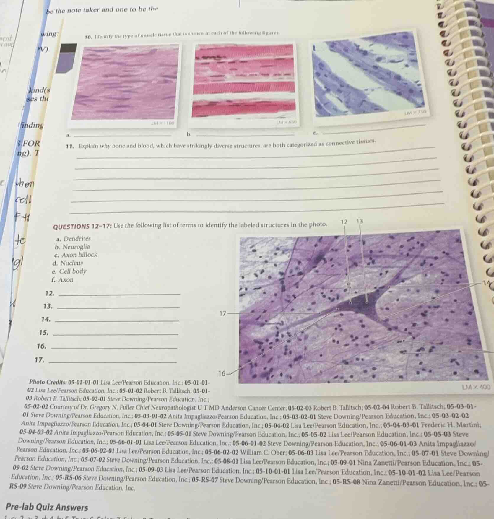
- identify the type of muscle tissue that is shown in each of the following figures.
a. \\_\\_\\_\\_\\_\\_\\_\\_\\_\\_\\_\\_\\_\\_\\_\\_\\_\\_\\_\\_\\_\\_\\_\\_\\_\\_\\_\\_\\_\\_\\_
b. \\_\\_\\_\\_\\_\\_\\_\\_\\_\\_\\_\\_\\_\\_\\_\\_\\_\\_\\_\\_\\_\\_\\_\\_\\_\\_\\_\\_\\_\\_\\_
c. \\_\\_\\_\\_\\_\\_\\_\\_\\_\\_\\_\\_\\_\\_\\_\\_\\_\\_\\_\\_\\_\\_\\_\\_\\_\\_\\_\\_\\_\\_\\_
- explain why bone and blood, which have strikingly diverse structures, are both categorized as connective tissues.
questions 12-17: use the following list of terms to identify the labeled structures in the photo.
a. dendrites
b. neuroglia
c. axon hillock
d. nucleus
e. cell body
f. axon
- \\_\\_\\_\\_\\_\\_\\_\\_\\_\\_\\_\\_\\_\\_\\_\\_\\_\\_\\_\\_\\_\\_\\_\\_\\_\\_\\_\\_\\_\\_\\_
- \\_\\_\\_\\_\\_\\_\\_\\_\\_\\_\\_\\_\\_\\_\\_\\_\\_\\_\\_\\_\\_\\_\\_\\_\\_\\_\\_\\_\\_\\_\\_
- \\_\\_\\_\\_\\_\\_\\_\\_\\_\\_\\_\\_\\_\\_\\_\\_\\_\\_\\_\\_\\_\\_\\_\\_\\_\\_\\_\\_\\_\\_\\_
- \\_\\_\\_\\_\\_\\_\\_\\_\\_\\_\\_\\_\\_\\_\\_\\_\\_\\_\\_\\_\\_\\_\\_\\_\\_\\_\\_\\_\\_\\_\\_
- \\_\\_\\_\\_\\_\\_\\_\\_\\_\\_\\_\\_\\_\\_\\_\\_\\_\\_\\_\\_\\_\\_\\_\\_\\_\\_\\_\\_\\_\\_\\_
- \\_\\_\\_\\_\\_\\_\\_\\_\\_\\_\\_\\_\\_\\_\\_\\_\\_\\_\\_\\_\\_\\_\\_\\_\\_\\_\\_\\_\\_\\_\\_
🆕 New Concept Discovered: Histological Identification of Tissues
Recognizing tissue types and cellular structures under a microscope.
Step 1: Identify Muscle Tissue Types (Question 10)
To identify the three types of muscle tissue shown in the micrographs:
- Image a: Shows striated, branching fibers with intercalated discs (dark bands connecting adjacent cells) and single, centrally located nuclei. This is characteristic of cardiac muscle tissue.
- Image b: Shows long, cylindrical, unbranched fibers with prominent transverse striations and multiple nuclei located at the periphery of the cells. This is characteristic of skeletal muscle tissue.
- Image c: Shows spindle-shaped (fusiform) cells that lack striations, each containing a single, centrally located nucleus. This is characteristic of smooth muscle tissue.
Step 2: Explain Connective Tissue Classification (Question 11)
Despite their vastly different physical structures and functions, bone and blood are both classified as connective tissues because they share three fundamental characteristics:
- Common Embryonic Origin: Both develop from mesenchyme (an embryonic tissue).
- Extracellular Matrix: Both consist of specialized cells suspended within an extracellular matrix (non-living material) rather than being tightly packed together.
- Structural Components: Both contain cells, ground substance, and fibers (even though the matrix of bone is calcified and solid, while the matrix of blood is liquid plasma with soluble fibers that only become visible during clotting).
Step 3: Identify Nervous Tissue Structures (Questions 12–17)
Using the provided list of terms to identify the labeled parts of the multipolar neuron and surrounding cells:
- 12 (Dendrites): The pointer indicates the highly branched, shorter processes extending from the cell body that receive incoming signals.
- 13 (Axon hillock): The pointer indicates the funnel- or cone-shaped region of the cell body where it tapers to connect to the axon.
- 14 (Axon): The pointer indicates the single, long, slender projection extending away from the cell body to transmit nerve impulses.
- 15 (Nucleus): The pointer indicates the large, round, centrally located organelle within the cell body of the neuron.
- 16 (Neuroglia): The pointer indicates the small, dark-staining nuclei of the supporting glial cells surrounding the large neuron.
- 17 (Cell body / Soma): The pointer indicates the main, expanded biosynthetic region of the neuron containing the nucleus and major organelles.
---

Snap & solve any problem in the app
Get step-by-step solutions on Sovi AI
Photo-based solutions with guided steps
Explore more problems and detailed explanations
10. Muscle Tissue Identification
- a. Cardiac muscle tissue
- b. Skeletal muscle tissue
- c. Smooth muscle tissue
11. Connective Tissue Explanation
Bone and blood are both categorized as connective tissues because they both originate from embryonic mesenchyme and consist of specialized cells widely separated by an extracellular matrix (consisting of ground substance and protein fibers).
Questions 12–17: Neuron Structure Identification
- 12. a. Dendrites
- 13. c. Axon hillock
- 14. f. Axon
- 15. d. Nucleus
- 16. b. Neuroglia
- 17. e. Cell body